


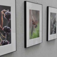
Linde Eyster enjoys looking closely at things — as a scientist, as a teacher, and as a photographer. For the past few years, she focused on the natural environment in her backyard garden, photographing a range of organisms with a macro lens. The result was a stunning, colorful collection that was on exhibition in Pieh Commons in October.
“I wanted the photos to tell biological stories,” says Linde, who has taught a variety of life science courses at Milton since 1990. “So, you’re not just looking at a photo of two ants. You are looking at a biological process. The ants are on a stem guiding the tiny aphids up and down, because the ants are dependent on the aphids for their nourishment.”
Linde shot all the images outside in natural light, with the subjects in their usual patterns and environment. The plan grew out of a cross-curricular biodiversity project she assigned her Advanced Biology students, who were required to find and photograph a dozen different invertebrates on campus or near their homes.
“I did the assignment myself to estimate how long it would take to accomplish, and the project reawakened my love of photographing little things,” says Linde. “Even without a camera in hand, I love the surprises of looking closely in the leaves and stems in my small garden, where I witness both amazing organisms and fascinating animal behaviors.”
Linde’s friend and fellow faculty member Bryan Cheney — a photographer and member of the visual arts department — answered her photography questions and helped her sort through hundreds of photos to select the ones to exhibit.
Linde’s interest in photography began as a child, when she occasionally converted the bathroom into a darkroom to develop her prints. For research toward her master’s degree, she took her first close-up photographs to document the colors of nudibranchs (sea slugs), which fade quickly when the organisms are placed into preservatives. During her doctoral research on embryonic shell formation at Northeastern University, she spent many hours photo-graphing subcellular structures with transmission electron microscopes, followed by hours of printing thousands of black-and-white images of cells and cell parts. Many of these images were published in her scientific papers.
“Without realizing it, I was learning skills through my microscope photography, such as composing the shot, finding the right angle, and cropping to focus on the elements you want,” says Linde.